22 Feb 2017

Unwinding the Double Helix: The Structure and Function of Nucleic Acids (Part 2)

The Three-Dimensional Structure of Nucleic Acids 

Nucleic acids, as with any molecule, possess a three-dimensional structure. Nucleic acids can be described into three main levels of structure: the primary, secondary, and tertiary structure. By describing these, we start to unravel why nucleic acids behave the way they do - and why they are capable of being the genetic material found in all forms of life and phages.

The Primary Structure of nucleic acids
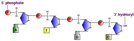
In the diagram above you can see that : the polynucleotide chain has a sense (directionality), where you 'read' it in the direction of the phosphodiester linkage between the 3' carbon of one monomer, to the 5' carbon of the next. The result is the nucleic acid carries an unreacted phosphate (Pi) group on the 5' end, and an unreacted 3' hydroxyl, the 5' to 3' logic is used to 'read' nucleic acids by ribosomes and other proteins involved with nucleic acid synthesis, replication etc. And as is the nature of nucleic acids, they each have a specific nucleotide sequence and for any one nucleic acid, this sequence is its primary structure.
When describing a polynucleotide, it is awkward and quite unnecessary to draw out the entire sequence of nucleotides, the diagram becomes cumbersome. There are several ways of representing the primary structure. For example, if you just want to represent the base sequence, you can do so like this (the first 40 nucleobases of human insulin, on chromosome 11) :
5'  ~[... AGCCCTCCAG GACAGGCTGC ATCAGAAGAG GCCATCAAGC ...]~  3'
In between each letter is a phosphodiester bond, all of these can be assumed to bond to be from a 3' hydroxyl to a 5' phosphate on the next nucleotide. Where the primary sequence ends with a 3',  is where the unreacted hydroxyl group is located; conversely, the 5' end is where the unreacted phosphate group occurs.

Hydrogen bonds and helices - the secondary & tertiary structure

The amazing helical shape that nucleic acids can form is familiar with most, it and similar structures occur due to the nucleobases interacting in respect to one another - this interaction results in the secondary structure. Some examples include the tRNA molecule and the double helix found in DNA. The secondary structure of DNA can vary in many forms (conformations) notably the A,B and Z forms. The majority of DNA adopts the B-DNA form, on the other hand, double-stranded RNA and DNA-RNA hybrids usually adopt the A conformation. These forms can occur due to changes in the chemical environment of the cell (including hydration level). You can see them below:
The tertiary structure on the other hand occurs only because of 'longer-range' interactions in the secondary structure. The best example of this occurs when DNA supercoils, compacting it and allowing the large molecule to fit inside cells and phage particles. Supercoiling is present in circular DNA and linear DNA. To understand how supercoiling works, first consider a B-DNA molecule that is base pairs in length; B-DNA typically has 10.5 base pairs per helical turns. Now that we know this we can consider this B-DNA molecule completes amount of turns, this number is what we call the Twist (T); if the circular DNA covalently joins at the last turn we say the molecule has a Linking number (L) of y. If this molecule. 
If we rotate this molecule counter-clockwise by one turn (360o), it becomes strained (as this reduces the Twist of the helix making it less stable), causing the B-DNA molecule to have 11.67 base pairs per turn (bp/turn). If the strained B-DNA molecule is allowed to return to its more stable conformation, it writhes (W) into a helix with 10.5 bp/turn once again. We can define the writhe as being negatively supercoiled, as W will have a value of -1. If the B-DNA molecule is rotated by two turns, W will have a value of -2, and so on. Overwound DNA is the opposite of this, by rotating it clockwise one turn W will be +1, and so on.
The degree of coiling in supercoiled DNA can be defined by the superhelix density (𝛔), which equals the change in linking number (𝚫L) over the linking number of the relaxed structure (L0), this relationship can be written as:
𝛔 = 𝚫L/L0
 Coiling also occurs in single-stranded nucleic acids whether they are RNA or DNA. For example: 
  • In denatured single strands there is considerable flexibility of nucleotides (residues), resulting in coils and no specific structure.
  • Non-self-complementary single strands, such as mRNA create a "stacked-base" structure, where the nucleobases stack, pulling the polynucleotide chain into a non-hydrogen bonding helix. The stacked-base structure is the normal shape assumed by these types of nucleic acid structure under physiological conditions.
  • Finally, the "hairpin" structure forms when single stranded nucleic acids are self-complementary. The strand folds back on itself, making a stem-loop structure, much like a hairpin. This can be seen in tRNA, an important molecule in gene expression. See below:

If you'be been reading my previous blogs, you would've noticed that the primary, secondary and tertiary structure of nucleic acids is somewhat analogous in definition to that of proteins. This is because both class of molecule share the same chemical principles when it comes to observing the structure of three-dimensional biological macromolecules. 

                                                                                                                             
Further reading:


As always, thanks for reading!

Don't forget to check out my Patreon,  if you like the content I'm putting out:


Links provided bring you to some of the info I used, the first year university textbook, "Biochemistry: Concepts and Connections," 1ED, by D.R. Appling, Pearson Ed. LTD, was used as a guide to write this post. You can buy it here

21 Feb 2017

Unwinding the Double Helix: The Structure and Function of Nucleic Acids (Part 1)

The Molecular Blueprints for Life


Depending on how long you've been following me for, you would know that I've already covered the basics of what DNA and RNA is, and how it works. In this series, we'll explore the what makes nucleic acids so special. For one, they are the only biological substance capable of self-replication thus enabling it to pass on information from one generation to the next. Inside this molecular code is information for everything that makes you, you - from the proteins that make your muscles, to the enzymes that allow you metabolise stuff. Mapped out and programmed, much of your development from child to adult, is coded within your very DNA. 

A Brief History of DNA

  • During the Franco-Prussian war in 1989, the military scientist Friedric Miescher 'discovered' DNA when he was analyzing discarded surgical dressings, to which he found very small quantities of some sort of acid. In this analysis, this acid was found predominantly in the nuclei of white blood cells.  Aptly, he named this substance "nuclein".
  • In the years 1884 to 1885, the scientists Oscar Hertwig, Albrecht von Kölliker, Eduard Strasburger, and August Weismann all provide evidence (independently) that the nucleus of a cell contain the information for inheritance.
  • Four years later, in 1889, Richard Altmann renames Miescher's discovery as "nucleic acids" instead of nuclein. 
  • By 1910, Thomas Hunt Morgan uses the Drosophila fruit fly to study inheritance, and discovers the white-eyed mutant. Three years later, with his colleague Alfred Sturtevant, they create the first genetic map for a chromosome in the Drosophila fruit fly. 
  • Frederick Griffith, in 1928, discovered that when a non-virulent strain of bacterium (Streptococcus pneumoniae) are able to become virulent when mixed with heat-disabled virulent strains. He called this the "transforming principle."
  • In 1929, Phoebus Levene discovers the building blocks of DNA.
  • In 1942, George Beadle and Edward Tatum make the discovery that genes are responsible for the production of proteins.
  • In 1944, Oswald T. Avery, Colin MacLeod, and Maclyn McCarty demonstrate that a discovery made in 1928 by Frederick Griffith is not the result of proteins being transferred between bacteria. Highly suggesting that nucleic acids are the genetic material. 
  • From 1949 to 1950, Erwin Chargaff discovers that the base composition discovered by Phoebus Levene varies between species in different quantities. 
  • Using the T2 bacteriophage, in 1952, Alfred Hershey and Martha Chase discover that it is the genetic material of virus, not the proteins that infect the bacterium. 
  • By 1953, Rosalind Franklin used x-ray diffraction to produce high-resolution images of the DNA structure, suggesting that it has a double-helix shape. Later that year, Francis Crick and James Watson produce the first model of DNA - a double helix in which the bases A always pairs with T, and C with G. Their discovery was published in April 25 1953 in the magazine Nature.
  • Matthew Meselson and Franklin Stahl discover how DNA replicates in 1958.
  • From 1961 to 1966, Robert W. Holley, Har Gobind Khorana, Heinrich Matthaei, Marshall W. Nirenberg and their colleagues manage to figure out what some genes code for what amino acids. "Cracking" the genetic code. 
  •  Paul Berg, in 1972 manages to create the first bit of recombinant DNA. By 1977, Frederick Sanger, Allan Maxam, and Walter Gilbert create the first method of sequencing DNA.
  • In 1982, the first commercial application of DNA technology using recombinant DNA is produced - human insulin becomes widely available and much easier to produce. A year later, Kary Mullis discovers the polymerase chain reaction (PCR) as a way to produce many copies of DNA in vitro.
  • After eight years, the sequencing of the human genome begins in 1990, which is only completed and then published 11 years later in 2001. The next year, the first genome of the model mammalian organism, the mouse, is completed.
The DNA molecule is recognised by everyone - the hallmark double helix structure is the go-to image for biosciences. With it, it has changed the way we think about the living world - it is the result of more than 100 years of hard work and dedication. Understanding DNA has lead us to develop new technologies to treat illnesses and hereditary diseases, and as demonstrated in 1982. The nature of how genes are passed on has been fundamental to modern agriculture and is responsible for the Green Revolution in the '30s to '60s and to now, where we are able to create hardier and more resilient crops than ever. You can check out my other blog series about Gene Editing here.

Two Types of Nucleic Acids

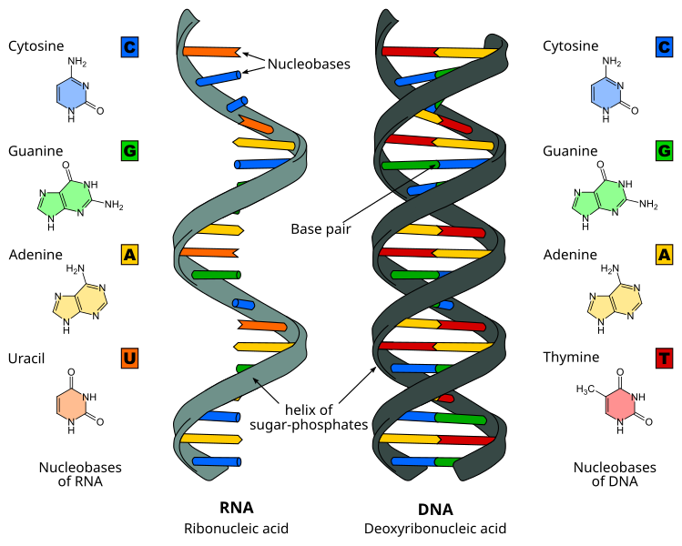
It is recognised that there are two types of nucleic acid: Miescher has discovered deoxyribonucleic acid (DNA), later it was discovered there was another type of nucleic acid, called ribonucleic acid (RNA). In each case, they are polymers - made of smaller molecules called monomers linked in a large chain. Their differences can be summarised below
Both RNA and DNA contain three main constituents which make up the nucleotides:
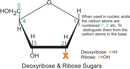
  • five carbon sugar, called ribose in RNA, and 2-deoxyribose in DNA. Their structure differs as below:
  • phosphate group, which forms a phosphodiester link between two sugar residues, forming the back bone of the nucleic acid. You can see how they look below:
  • And finally, the nucleobases, which can be seen above and are bonded to the 1' carbon of the sugar. These bases form hydrogen bonds with the adjacent strand, and in the case of mRNA, pair with tRNA to enable protein production. They can be separated into two types: the purines (Adenine and Guanine) and the pyrimidines (Cytosine (and 5-methylcytosine) ,Thymine, and Uracil which is found RNA only). The chemical bond between the carbon 1' of the sugar and the nucleobase is termed as a glycosidic bond.

Nucleotides and their derivatives

As noted above, nucleotides are the monomers that make up the DNA strand and are connected via phosphodiester linkages. Nucleotides are considered to be the phosphorylated derivative of a nucleoside, which lack the phosphate group on the 5' carbon of the sugar group. Large stretches of nucleotides are often called polynucleotides while those with a few are termed oligonucleotides (dinucleotides, trinucleotides, tetranucleotides etc.).
Noted before as well, RNA contains the ribose sugar, in which the 2' OH group is still present, giving it a different function than with the DNA deoxyribose form. This is because the 2' OH group is found in RNA enzymes called ribozymes, which were discovered by Thomas Cech and Sidney Altmann independently. Because of this, many biochemists think that it is possible that RNA came into existence earlier than DNA. DNA is much more stable than RNA, which allows it to be much larger than its ribose counterpart. 
Nucleotides are strong acids where the ionization of the phosphate group and the deprotonation/protonation of the bases at pH values around 7. The nucleobases are also capable of converting into different tautomeric forms because of the several double bonds present in the ring structure (a form of conjugation). For example, uracil can convert between the keto and enol forms:
A consequence of highly conjugated chemical structures is how much the molecules absorb light. Purines, pyrimidines and their derivatives (nucleic acids, nucleotides and nucleosides) all absorb light in the ultraviolet region. You can scroll down on the papers here, and here to see these spectra. Normally we use these spectra to make quantitative measurements at the 260 nm setting on a spectrophotometer
Another important part of nucleotides are the phosphodiester bonds present in the DNA molecule. These bonds are formed by "adding" a water to the monomers. The opposite is true, hydrolysis removes a water molecule from this bond, releasing a ∆Gº' = +25 kJ/mol, as a result, this is a thermodynamically favoured reaction. See below to see how this looks like:
In the cell, both RNA and DNA are broken down by nucleases, catalysing reaction (2) above, generally this reaction is utilised in metabolic pathways as a way to create glucose, or ketone bodies to produce energy, excess nitrogen is converted into urea and then excreted (more on this in my Metabolism and the Energy of Life series). As discussed in that series, ATP is often used by the body to drive forward reactions that would otherwise be impossible in vivo. The polymerization of nucleotides is such a reaction, leading to the phosphodiester bond's stability.
In fact, DNA is so stable, it has been found in bones recovered to be as old as 80,000 years old. This assisted us in sequencing the complete genome of an extinct human species, the Neanderthal in 2010.  The resilience of DNA has also helped us map the genetic distribution of our own species, from Africa to beyond. A truly wonderful molecule!


Now that you've been acquainted with nucleic acids. the next section will go over its primary structure. 
As always, thanks for reading! 
Don't forget to check out my Patreon,  if you like the content I'm putting out:

Links provided bring you to some of the info I used, the first year university textbook, "Biochemistry: Concepts and Connections," 1ED, by D.R. Appling, Pearson Ed. LTD, was used as a guide to write this post. You can buy it here

20 Feb 2017

Your body's defense system: Antibodies (Part 3 of 3)


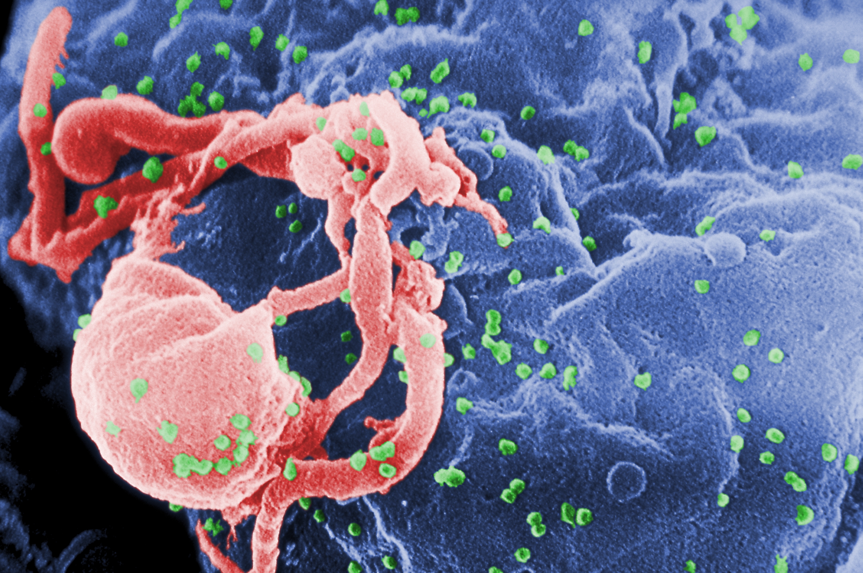
The above image is a colourised electron micrograph that of a T-cell  (green) infected by HIV H9 viruses (yellow) budding on the cellular surface. It would be beautiful, were it not so deadly.
                                                                                                                                

 Antibodies are used in an array of different defence situations

So far, I've only covered how your immune system produces antibodies that tag and aggregate an antigen (foreign substance) which is then consumed by a macrophage (white blood cell) in a process known as the humoral immune response. But there's the other form of defense too: the cellular immune response. As we've established previously, the cellular immune response involves a specific set of cells called lymphocytes that recognise and destroy foreign cells (antigens).
When people undergo transplants, it's commonplace that a few patients' bodies will undergo tissue rejection. The cellular immune response plays a role in this phenomenon as cells that don't quite match you own have different receptors on the cell surface, this leads to lymphocytes rejecting the donor's organ as those regions are recognised as an antigen. Your body also can utilise this system to destroy any potential cancer cells before they propagate to unmanageable quantities, this doesn't always work, though.
In both humoral and cellular immune responses have a similar molecular method of recognising foreign objects by using proteins belonging to the immunoglobulin superfamily. In the four examples below, if your eyes are good, you'll notice they all share similar sheet and loop structures. PDB IDs (protein database ID) are and links to their RCSB PDB profiles are listed:

Left: A class I human major histocompatibility complex (MHC), PDB ID: 1a1mRight:  A class II human MHC, PDB ID: 1dlh


Left: A human T-cell receptor binding to a MHC class I molecule and a viral peptide; PDB ID: 1bd2Right: A Murine Fab  fragment; PDB ID: 1nca (the link shows for Fab fragments bound to a neuraminidase.
I'd definitely recommend checking out those RCSB links; they have 3D models of these molecules so you can have some idea of how they bind to antigens (some of the models include an antigen).
In the header and throughout this part I've mentioned T cells quite a lot. These cells are involved in the cellular immune response and have structures similar to the Fab  fragments of antibodies on their cellular surface. Consequently, these cells target unwanted cells and are able to destroy them, which is why they have the name of Killer T cells ( or "cytotoxic T cells"). Receptors on the outside of these cells are capable of recognising foreign peptide chains on the surface of invading or infected cells. The examples shown above include MHC proteins, these are found on killer T cells and act as a switch that releases an attack protein called perforin. Perforins are released only when a T cell receptor (including the MHCs) detect an antigen, these then react with the detected invasive cell's membrane and form pores that effectively drain the cell of essential ions - killing the cell.

Why AIDS vaccines are so hard to produce based on what we've covered so far

Acquired Immune deficiency syndrome (AIDS) is a symptom of the problematic human immunodeficiency virus (HIV). HIV attacks T cells that are essential in a healthy immune system. The infected type of T cells that HIV infects are part of the first line of defence against antigens - they signal a set of B cells that produce important antibodies that help identify and destroy foreign objects. In effect, HIV completely disables the body's ability to defend itself.
To make matters worse, HIV undergoes mutations in it's genome (and therefore the antigenic determinants change often) at 60 times the rate of the influenza virus. Making a flu vaccine is problematic enough because the same reason - once we make a vaccine, and many viruses are destroyed, those remaining (somewhere in the human population) have already mutated and cannot be recognised by a complementary antibody.
So far we've only been able to slow the progress of AIDS, through therapies involving drugs that target the processes that replicate the viral genome in cells.
Here's hoping some brilliant person will figure out a way to stop the AIDS pandemic which has ruined 60 million lives since 1983.

                                                                                               
And that concludes this short three-part series! Make sure to look into more of this subject.

Your body's defense system: Antibodies (Part 2 of 3)

E coli bacterium


Pictured above is the E. coli bacteria. Some strains of these are pathogenic, others (found in your digestive system) aren't dangerous at all.
                                                                                                                              

Immunoglobulin structure, function and diversity

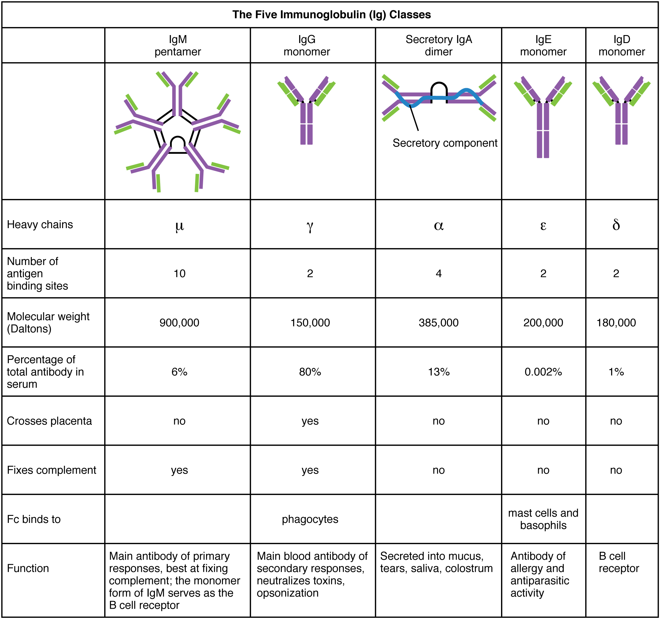
There are five classes of immunoglobulin (antibody) molecules found in your body's arsenal - they all share a common basic structure which can be seen in other mammals' antibodies too. Every antibody is consists of one to five different immunoglobulins; when there is more than one immuoglobulin present, they are linked together by a molecule called a J chain. Antibodies are large molecules that we call a polymer as it is made of smaller units called monomers, in this case they are in the form of immunoglobulin - joining together to make a larger antibody unit*.
*To clarify - in the previous section I said immunoglobulin and antibody are interchangeable, this is still true as individual immunoglobulin molecules  join together (as each has a different function) and form what we call an antibody. Being made of several differnt immunoglobulin monomers gives the antibody versatility when engaging threats. The different types of a immunoglobulin monomers are below:
The immunoglobulin monomer is made of four polypeptide chains and can be separated into two forms: two heavy chains with a molecular weight of 53,000 Da (Daltons) and two light chains with a molecular weight of 23,000 Da each. Each of these chains are held together by Disulfide bonds (S-S) and contain regions of called domains. These domains are either constant (the same in any antibody of a type of class) or variable.
The variable domains of these chains determine how the antibody binds to a specific antigen (foreign object). Large objects, such as a protein, virus or bacterial cell have different parts that can be recognised by an antibody (antigenic determinants), these areas are usually scattered on the object's surface, allowing more than one antibody to bind and force the antigen to aggregate. This type of aggregation is called immunoprecipitation - and works by isolating a threat to be destroyed that contains thousands of different proteins or antigenic determinants. Immoprecipitation is a process that requires an antibody to have a bivalent structure, meaning it needs two areas in which it can bind to the antigen. 
In the lab, it's possible to break up the antibody into fragments ("cleavage"); the moleucle is of say, IgG is Y shaped and produces three fragments: two Fabfragments contains a binding site each) and a single Fc fragment (contains no binding sites for antigens). See the image below, noting that V denotes a variable chain and C denotes a constant chain. It's also important to note that the  innermost chains of the Y shape are heavy chains while the outermost chains are the light chains:
The constant domains (C) help keep chains together and also act as signalling regions (effectors) to other cells involved in immune responses such as T-cells or macrophages (white blood cells). 

The artful nature of antibodies: the immunoglobulin superfamily

Domain areas in immunoglobulins have a common canvas called the Ig domain which most likely presents a primitive structural element found in the evolution of the immune response. Proteins that have this Ig domain are classed as being part of the immuoglobulin superfamily. This domain is a very stable scaffold which hold the hypervariable molecular loops which determine the shape and charge of the areas that bind to antigens.  The molecular loops are called complementary determining regions (CDRs).
The CDRs are what determines whether or not the antibody will bind to an antigen, this is due to the  shape and charge complementarity  of the CDRs. Shape complementarity occurs because the three-dimensional shape of the antibody binding area and the shape of the antigen complement each other and fit like a puzzle piece or say, a glove. Charge complementarity on the other hand occurs when weak interactions between the target antigen and antibody, such as van der Waals, hydrogen bonding and electrostatic attractions. These interactions are the same as explained in my other blog here. These types of interactions are useful in biochemistry as they help explain many different types of biochemical processes, such as the structure of DNA.
It is our understanding of the molecular binding between the CDRs and the antigens that helps efforts to create vaccines against the deadly hepatitis B - affecting around 400 million people worldwide. 

Generating Antibody diversity

Throughout life's time here on earth, particularly in large animals such as ourselves and our ancestors, B-cells that produce antibodies have undergone many mutation events such as sequence rearrangement or splicing that have resulted in numerous combinations of genetic code. The genetic code for IgG immunoglobulin (in the table) CDR loops has historically mutated at an unusually high rate in mammals and accounts for the diversity of combinations of IgG immunoglobulins found in the human genome (there's around 10 billion combinations!). The mutation events are random and are therefore not pre-programmed thus it is possible for a white blood cell (B lymphocyte) to produce an immune response to synthetic substances that have a binding sites complementary to the antibodies.

                                                                                                                               
The next section will cover some cellular immune response cells involved in producing antibodies such as killer T cells and why it's so hard to develop a vaccine for AIDS.
Thanks for reading! 

19 Feb 2017

Your body's defense system: Antibodies (Part 1 of 3)

Immunoglobulins: Part of your cellular army


Immunoglobulins, otherwise known as antibodies are large proteins, that come in a wide range different types; but they all come with a similar structural framework. Antibodies bind to a target with discriminate specificity - causing irreversible damage to the target antigen (foreign object within the body, ranging from viruses, bacteria or a type of molecule).
The body's immune response is highly adaptive and is able to detect a foreign substance, and so defends itself via an (adaptive) immune response. This response comes in two forms: the humoral and cellular immune responses. 
Humoral responses involve lymphatic cells called B lymphocytes that produce cells that act as a foreign substance tagging system - they bind to the target antigen, causing the foreign substance to group together, and thus letting the body know that it needs to be destroyed. The cells that detect and destroy antigens that are tagged are called macrophages. T lymphocytes involved in the cellular immune response locate and destroy foreign cells.
Provided the antigen is large enough (like a virus, bacterial cell or a protein), more than one type of antibody may be produced, since substances that are large generally have different components that require different antibodies to bind to, and destroy the foreign object.
The body's immune response is highly adaptive, but it's even capable of having a sort-of "memory" system. For example, an initial antigen is encountered, the body produces antibodies to combat the invading substance. The next time the substance enters the body, the immune response will be even quicker and even producing more antibodies than the previous time.
Vaccines are developed with this in mind - pathogens that have been modified with a significantly reduced ability to cause disease, but still contain the antigen properties required for the body to illicit an immune response. Antibodies are produced in the body which destroy the antigen, memory cells are able to produce more antibodies if the foreign substance is encountered again.
Immature B lymphocytes are produced within the bone marrow, every cell produces one immunoglobulin molecule which are able to recognise a specific antigen because different molecules have different shapes - the antibodies have binding sites that recognise these shapes. Antibodies that do this are located on the outer membrane of B lymphocytes, these cells are constantly circulating in your bloodstream. 
With the help of T helper cells, the B lymphocytes are able to produce antibodies that are soluble (mixes with the water in your bloodstream) which are able to move around the bloodstream freely. This response called is the Primary immune response and is carried out by a type of B lymphocyte called plasma cells (or, Effector B cells). The other type of B lymphocyte are memory cells, which stick around for quite a while, allowing for a rapid secondary immune response if the antigen is encountered again.
Sometimes, there is an error in immature B lymphocytes produced in bone marrow, and they accidentally bind to tissues that are part of the body - this problem is called autoimmunity, and the reasons for this are still quite unclear.
Knowing how the body manages its defense systems is vital in medicine today - it not only provides us with the means to produce more effective vaccines, but it also allows us to map out the genetic history of antibodies. 

18 Feb 2017

Chemical Wonders: Experimental Techniques (Part 3) - Chemistry in the Infrared

Infrared & Raman Spectroscopies


Infrared spectroscopy is an invaluable tool for chemists, and is applied in fields from astronomy to forensics. It takes advantage of the transitions between vibrational energy states of molecules, and in order to be classed a 'IR active,' there need to be a change in the charges of the dipole moment. The other mode of vibration, called Raman spectroscopy, deals specifically with the change in polarizability. Both IR and Raman spectroscopy are forms of vibrational spectroscopy. The IR region ranges from 20 cm-1 to 14 000 cm-1 (called near IR).

The wavenumbers of molecular vibrations

When a molecule is exposed to infrared radiation, its covalent bonds vibrate and stretch (think of it like stretching a spring), when this occurs the molecule undergoes harmonic oscillations. The energy levels of these vibrations are given by:
Ev = (v + ½)hv - (v + ½)2 hvxe      (Ev is in J, joules)
where v = vibrational quantum number; h = Planck constant; v = frequency of vibration; x = anharmonicity constant. At an energy level, where v = 0, is the zero point energy of the molecule. When dealing with a transition from the vibrational ground state, to the first excited state, the motion of this molecule is approximately one of a simple harmonic oscillator:
Ev = (v + ½)hv 
When considering a hypothetical diatomic molecule, say, XY you would find that the vibrational frequency is dependant on two factors: the mass of atoms X and Y; and the force constant (k) of the bond. The constant, k, is determined by the strength of the covalent bond or in other words the stiffness of the "spring." 
  • Diatomic molecules with where X and Y similar masses, they approximately contribute equally to the molecular vibration.
  • In molecules where X and Y have significantly different masses, the lighter atom moves more than the heavier one.
The reduced mass,𝜇, is the quantity that describes the mass of the oscillator so that it can more accurately reflects the relative masses of X and Y. This relationship is given by:
1/𝜇 = 1/mx + 1/my           OR           𝜇 = mxmy / mx + my 
The fundamental absorption of the molecule is the transition from the ground state to the first excited state. This relationship can be given by:
v = 1/2𝜋 √k/𝜇
where: v = the fundamental vibrational frequency (Hz); k = force constant (N m-1);  𝜇 = reduced mass; 𝜇 = reduced mass. Using these definitions, we can define the relationship of the absorptions in IR spectra in relation to the wavenumber, it can be defined as below:
 \scriptstyle\tilde{\nu}= 1/2𝜋c√k/𝜇 
where  \scriptstyle\tilde{\nu}wavenumber (cm-1); c = speed of light  = 3.00x1010 cm s-1.

Characteristics of IR spectra

The IR spectra produced from using a device such as the Fourier transform infrared spectrometer (FT-IR, shown above), can be separated into two main regions: the fingerprint region, which occurs in bands in the regions below 1500 cm-1, which arise from single bond stretching nodes, vibrations within the molecule and deformations in the molecular structure. Any absorption wavelength found above this region is typically what is used to identify key functional groups on the compound in question. The fingerprint region on the other hand is used to identify the characteristic signature of the compound, as the bands in these regions are specific to the overall structure of the molecule.
You can find a table of these functional groups here.
I mentioned that the IR spectra ranges from 20 cm-1 to 14 000 cm-1 (called near IR), however, in the lab, IR spectrometers typically use the range from 400 to 4000 cm-1, dubbed the 'mid-IR' section of the spectra. Data produced from the machines, like the FT-IR produce spectra in which the transmission (at arbitrary values 0 to 100%) against the wavelength of the IR bands. Samples in a typical FT-IR machine can be recorded using samples in gaseous, liquid or even solid samples. Samples in different states need to be prepared in different ways and result in slightly different IR spectra. 
Solids are typically prepared in a mull, mixing the solid with an organic oil, or it is pressed into a disc by grounding it with a an alkali metal halide (such as KBr). These forms of preparation affect the IR spectrum: the disc preparation reduces the range observed by the spectra, as it is transparent from 4000 to 450 cm-1, while NaCl is from 4000 to 650 cm-1. More modern machines utilise diamonds, the accessory known as a diamond attenuated total reflector (ATR), which allows us to avoid using mulls or discs.

Raman Spectroscopy

IR and Raman spectroscopy are two techniques which can be used together, and in 1930 Chandrasekhara V. Raman (pictured left) won the noble prize in physics. He discovered that radiation is scattered when a molecule is exposed to a frequency, v0, even though there is no change in frequency. This is called Rayleigh scattering, which is also responsible for the blue colour of the sky. A small amount of scattered radiation has frequencies of v0 ± v, where v is frequency of the vibrating section of the molecule. This is known as Raman scattering. It is actually quite an unsensitive because a only a small range actually undergoes Raman scattering. Improvements have been made by using Fourier transformation (FT) techniques. 
Raman spectroscopy is particularly useful because it utilises wavelengths below the normal IR spectroscopy range, this in turn, allows chemists to observe the vibrational nodes found in metal-ligand bonds. Coloured compounds rely on laser excitation that coincide with the absorption wavelengths in the electronic spectrum, known as resonance Raman spectroscopy. Utilising resonance enhancement allows for more clearly defined lines.

________________________________________________________

Don't forget to check out my Patreon,  if you like the content I'm putting out:


Links provided bring you to some of the info I used from the web. the first/second-year university textbooks:
  • "Inorganic Chemistry," 4ED, by Housecroft & Sharpe, Pearson Ed. Ltd., 'Chapter 4 - Experimental techniques,' pgs 90-98. You can buy it here.
  • "Fundamentals of Analytical Chemistry," 9ED, Skoog & West, Pearson Ed. Ltd., 'Chapter 28 - Atomic Spectroscopy,' pgs 774-775, 790-799. You can buy it here.

17 Feb 2017

Chemical Wonders: Experimental Techniques (Part 2) - Identifying Elements and Ions in a Compound

When identifying compounds in a lab, you utilise different techniques to get an accurate data output for the compound in question. This part will focus two important parts of experimental chemistry (you may recognise some things from high school chemistry): elemental analysis using techniques such as combustion, atomic spectroscopy, and mass spectrometry, which utilises the ionization of the molecule.

Mass Spectrometry (MS): Separating ions


There are a variety of mass spectrometry techniques which all share one thing in common: separating ions from the compound, whether they are atomic or molecular, and separating them according to their mass-to-charge ratio (m/z). I'll only cover two classic techniques, such as Electron Ionization (EI-MS), and electrospray (ESI) techniques. Other techniques include: fast atom bombardment (FAB) and as well as matrix assisted laser desportion ionization time-of-flight (MALDI-TOF). These techniques are familiar to all chemists and biochemists as extensively useful tools in analytical chemistry,

Electron Ionization (EI) Mass Spectrometry

Electron Ionization or electron impact mass spectrometry would probably be the most familiar to you if you did high school chemistry. This technique is known as a 'hard technique' as it involves bombarding the analyte with high-energy electrons (  70 eV, electron volts), causing the molecule to fragment thus producing ions. It is widely used for analysing organic compounds, but becomes increasingly more limited as the molecular mass of a compound increases (Mr < 1500). This technique can't be used on ionic compounds, and the analyte must be stable when vaporized (if it isn't a gas at 298.15 K). As such this technique is limited to vaporized substances. In general, the molecular breakage can be represented generally as:
M(g) + e-(high energy) -------------> [M]+ (g) + 2e- (low energy)
Using a high-energy electron stream is essential because it is used to break the high covalent bond energies present in the analyte. Two ions are produced, the parent ion and the ion. The [M]+ (g) cation is a radical and is written as [M]which are highly reactive. Fragmentation of molecules is always considered a 'hard technique.' After bombardment with electrons, they pass through a magnetic field, where the positive ions are deflected into a detector. Deflection is entirely dependent on the size of the m/z ratio: a larger m/z value leads to a greater radius of deflection. For ions with a value of z = 1, the m/z value is the same as the molecular mass; if z=2, the m/z ratio is half that of the molecular mass of the ion, and so on.
However, due to being restricted to molecules with relatively low molecular and low energy of vaporization, most ions have z=1. The mass spectrum is plotted so that m/z lies on the x-axis, and the y-axis is the relative intensity of the fragments, arbitrarily set on a scale of 0 to 100%. The final output depends on isotopes of elements that may be present in the molecule too, leading to an observation termed peak envelopes. The device can be summarised below:

Electrospray Ionization (ESI) Mass Spectrometry

This technique is widely used in molecules that have relatively high molecular weight (Mr ≤ 200 000). In contrast with EI mass spectrometry, this technique can be used with ionic substances, where singly and multiply charged ions can be observed in the resultant mass spectrum. This gives give it an advantage over the EI technique. It is also termed a 'soft' technique, as it involves the injection of a sample dissolved in a volatile (such as MeCN or MeOH which are easily vaporized) solvent, which is then sprayed (at 1 atm) into an applied electrical potential. The potential between the original point of injection to the counter electrodes is ~3000 V in positive ion mode. As the ions move toward the counter electrodes, the solvent evaporates and  the gas-phase ions produced eventually hit the mass analyser. Peaks produced that are one mass unit apart reveal an ion is singly charged; on the other hand if they are half a mass unit apart, it is doubly charged, and so forth.
Like FAB and MALDI-TOF, neutral molecules are converted into positive ions with the help of H+ and Na+. As a result, an aggregate may be produced, generally they either result in a [2M+Na]+ and  [M + MeCN + H]+.

Elemental and Compositional Analysis

Combustion

For a quantitative analysis of carbon, hydrogen and nitrogen containing compounds, it is possible to fully combust the compound and using the reaction stoichiometry, reach a conclusion for the composition of an analyte. It is done when a known mass of a substance (e.g. 2 to 5 mg) is sealed in an aluminium or tin capsule. This is placed in a fully automated analyser, where it is injected into a pyrolysis/combustion tube and heated to 900 ℃ in a pure oxygen environment. For compounds containing C, H, and N, they are oxidised into CO2, H2O and nitro-oxide gases, respectively.
These are then moved using a carrier gas (He) into a copper chamber, where nitro-oxides are reduced into N2 gas and excess O2 is removed. From here, the CO2 and H2O gas mixture is separated and then moved into and analysis chamber, where separated using a type of gas chromatography, which doesn't have a mobile phase (refer to the previous part of the series). The separated gases are then detected using a thermal conductivity detector, the detection process takes about five minutes. The accuracy the recorded amounts of C, H and N present in the compound is about <0.3%.
More modern machines can determine the amount of O and S present, where they are converted into SO2 and CO to CO2, respectively.    

Atomic Absorption Spectroscopy (AAS)

This technique is used for determining the quantitative amount of metal but utilising the absorption and emission spectrum of elements. For example, the emission spectrum of hydrogen consists of very sharp lines, each of which corresponds to electronic transitions between high/low energy levels. On the other hand, the absorption spectrum of hydrogen occurs when it becomes irradiated, each element has its own absorption and emission spectrum. AAS is a common type of spectroscopy which uses a hollow cathode lamp calibrated to a given wavelength (specific to a transmission from one energy state to another), which irradiates the analyte. 
Generally, the metal being analysed isn't present in its pure elemental form, so the analyte must be broken down (a step called digestion) in a series of standards (in liquid form). Standards are used to construct calibration curve. Each standard passes through a nebulizing chamber, where oxygen is injected in the liquid sample, resulting in very fine spray. The spray then enters an atomizer (generally a flame atomizer/graphite furnace/electrically heated), where the sample becomes atomized. The hollow cathode lamp irradiates the atomized sample, which passes through the monochromator. This is an optical component that transmits a beam of light with a very narrow range (basically a single colour), by reflecting away the unwanted wavelengths. The results are then amplified and the absorption spectrum is output to a computer display.
Modern AAS devices are computer-controlled, where the data is automatically recorded processed onto the computer. The AAS device is extremely sensitive - in the range of μg dm-3 ! 

_______________________________________
As always, thanks for reading! 

Don't forget to check out my Patreon,  if you like the content I'm putting out:


Links provided bring you to some of the info I used from the web. the first/second-year university textbooks:
  • "Inorganic Chemistry," 4ED, by Housecroft & Sharpe, Pearson Ed. Ltd., 'Chapter 4 - Experimental techniques,' pgs 90-98. You can buy it here.
  • "Fundamentals of Analytical Chemistry," 9ED, Skoog & West, Pearson Ed. Ltd., 'Chapter 28 - Atomic Spectroscopy,' pgs 774-775, 790-799. You can buy it here.