Showing posts with label educational. Show all posts

20 Feb 2017

Your body's defense system: Antibodies (Part 3 of 3)


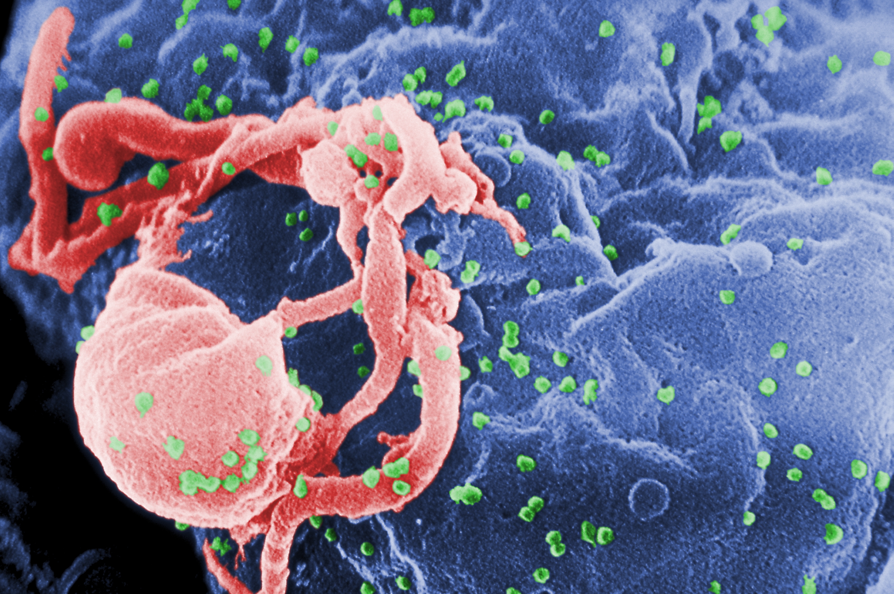
The above image is a colourised electron micrograph that of a T-cell  (green) infected by HIV H9 viruses (yellow) budding on the cellular surface. It would be beautiful, were it not so deadly.
                                                                                                                                

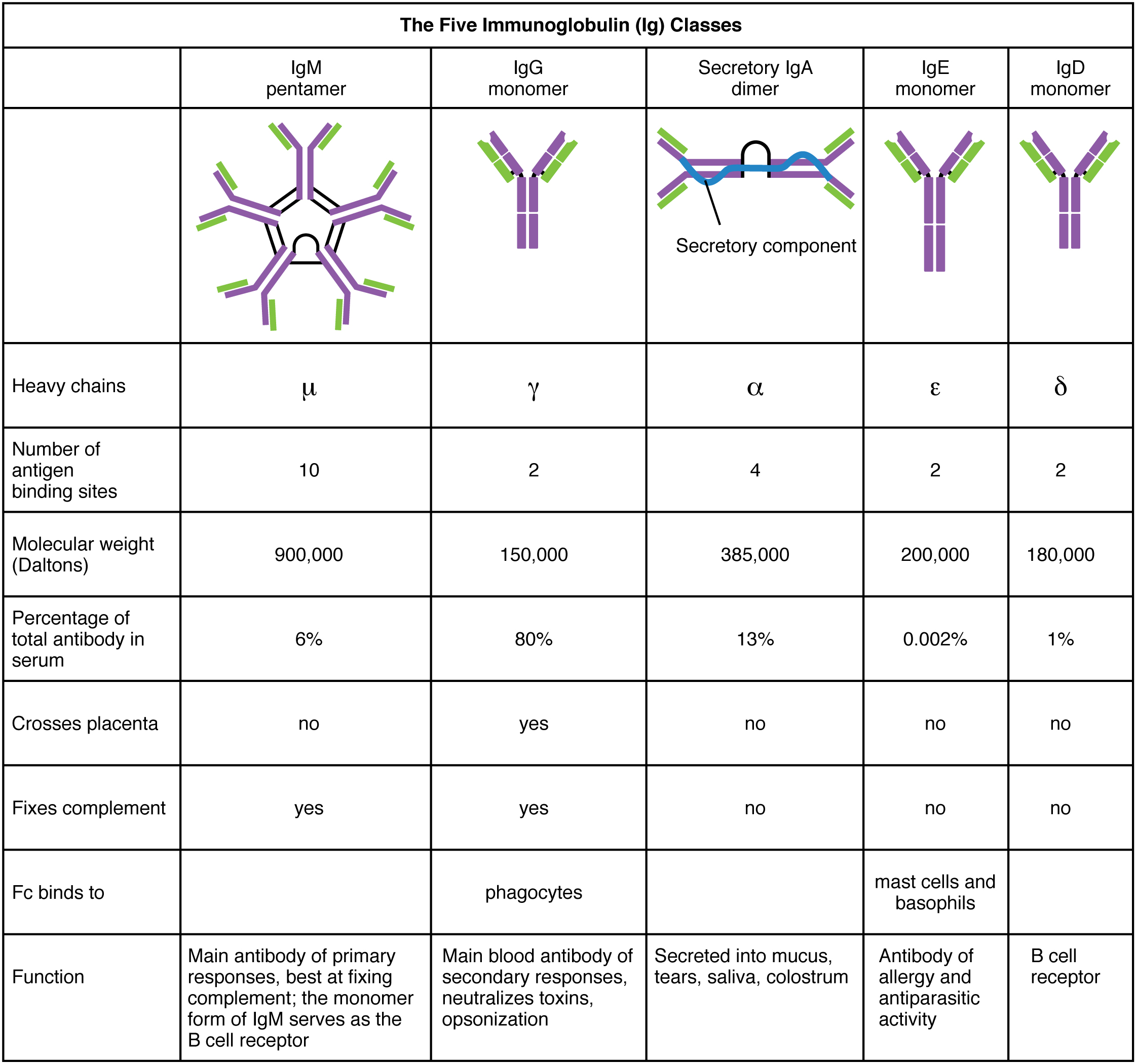
 Antibodies are used in an array of different defence situations

So far, I've only covered how your immune system produces antibodies that tag and aggregate an antigen (foreign substance) which is then consumed by a macrophage (white blood cell) in a process known as the humoral immune response. But there's the other form of defense too: the cellular immune response. As we've established previously, the cellular immune response involves a specific set of cells called lymphocytes that recognise and destroy foreign cells (antigens).
When people undergo transplants, it's commonplace that a few patients' bodies will undergo tissue rejection. The cellular immune response plays a role in this phenomenon as cells that don't quite match you own have different receptors on the cell surface, this leads to lymphocytes rejecting the donor's organ as those regions are recognised as an antigen. Your body also can utilise this system to destroy any potential cancer cells before they propagate to unmanageable quantities, this doesn't always work, though.
In both humoral and cellular immune responses have a similar molecular method of recognising foreign objects by using proteins belonging to the immunoglobulin superfamily. In the four examples below, if your eyes are good, you'll notice they all share similar sheet and loop structures. PDB IDs (protein database ID) are and links to their RCSB PDB profiles are listed:

Left: A class I human major histocompatibility complex (MHC), PDB ID: 1a1mRight:  A class II human MHC, PDB ID: 1dlh


Left: A human T-cell receptor binding to a MHC class I molecule and a viral peptide; PDB ID: 1bd2Right: A Murine Fab  fragment; PDB ID: 1nca (the link shows for Fab fragments bound to a neuraminidase.
I'd definitely recommend checking out those RCSB links; they have 3D models of these molecules so you can have some idea of how they bind to antigens (some of the models include an antigen).
In the header and throughout this part I've mentioned T cells quite a lot. These cells are involved in the cellular immune response and have structures similar to the Fab  fragments of antibodies on their cellular surface. Consequently, these cells target unwanted cells and are able to destroy them, which is why they have the name of Killer T cells ( or "cytotoxic T cells"). Receptors on the outside of these cells are capable of recognising foreign peptide chains on the surface of invading or infected cells. The examples shown above include MHC proteins, these are found on killer T cells and act as a switch that releases an attack protein called perforin. Perforins are released only when a T cell receptor (including the MHCs) detect an antigen, these then react with the detected invasive cell's membrane and form pores that effectively drain the cell of essential ions - killing the cell.

Why AIDS vaccines are so hard to produce based on what we've covered so far

Acquired Immune deficiency syndrome (AIDS) is a symptom of the problematic human immunodeficiency virus (HIV). HIV attacks T cells that are essential in a healthy immune system. The infected type of T cells that HIV infects are part of the first line of defence against antigens - they signal a set of B cells that produce important antibodies that help identify and destroy foreign objects. In effect, HIV completely disables the body's ability to defend itself.
To make matters worse, HIV undergoes mutations in it's genome (and therefore the antigenic determinants change often) at 60 times the rate of the influenza virus. Making a flu vaccine is problematic enough because the same reason - once we make a vaccine, and many viruses are destroyed, those remaining (somewhere in the human population) have already mutated and cannot be recognised by a complementary antibody.
So far we've only been able to slow the progress of AIDS, through therapies involving drugs that target the processes that replicate the viral genome in cells.
Here's hoping some brilliant person will figure out a way to stop the AIDS pandemic which has ruined 60 million lives since 1983.

                                                                                               
And that concludes this short three-part series! Make sure to look into more of this subject.

Your body's defense system: Antibodies (Part 2 of 3)

E coli bacterium


Pictured above is the E. coli bacteria. Some strains of these are pathogenic, others (found in your digestive system) aren't dangerous at all.
                                                                                                                              

Immunoglobulin structure, function and diversity

There are five classes of immunoglobulin (antibody) molecules found in your body's arsenal - they all share a common basic structure which can be seen in other mammals' antibodies too. Every antibody is consists of one to five different immunoglobulins; when there is more than one immuoglobulin present, they are linked together by a molecule called a J chain. Antibodies are large molecules that we call a polymer as it is made of smaller units called monomers, in this case they are in the form of immunoglobulin - joining together to make a larger antibody unit*.
*To clarify - in the previous section I said immunoglobulin and antibody are interchangeable, this is still true as individual immunoglobulin molecules  join together (as each has a different function) and form what we call an antibody. Being made of several differnt immunoglobulin monomers gives the antibody versatility when engaging threats. The different types of a immunoglobulin monomers are below:
The immunoglobulin monomer is made of four polypeptide chains and can be separated into two forms: two heavy chains with a molecular weight of 53,000 Da (Daltons) and two light chains with a molecular weight of 23,000 Da each. Each of these chains are held together by Disulfide bonds (S-S) and contain regions of called domains. These domains are either constant (the same in any antibody of a type of class) or variable.
The variable domains of these chains determine how the antibody binds to a specific antigen (foreign object). Large objects, such as a protein, virus or bacterial cell have different parts that can be recognised by an antibody (antigenic determinants), these areas are usually scattered on the object's surface, allowing more than one antibody to bind and force the antigen to aggregate. This type of aggregation is called immunoprecipitation - and works by isolating a threat to be destroyed that contains thousands of different proteins or antigenic determinants. Immoprecipitation is a process that requires an antibody to have a bivalent structure, meaning it needs two areas in which it can bind to the antigen. 
In the lab, it's possible to break up the antibody into fragments ("cleavage"); the moleucle is of say, IgG is Y shaped and produces three fragments: two Fabfragments contains a binding site each) and a single Fc fragment (contains no binding sites for antigens). See the image below, noting that V denotes a variable chain and C denotes a constant chain. It's also important to note that the  innermost chains of the Y shape are heavy chains while the outermost chains are the light chains:
The constant domains (C) help keep chains together and also act as signalling regions (effectors) to other cells involved in immune responses such as T-cells or macrophages (white blood cells). 

The artful nature of antibodies: the immunoglobulin superfamily

Domain areas in immunoglobulins have a common canvas called the Ig domain which most likely presents a primitive structural element found in the evolution of the immune response. Proteins that have this Ig domain are classed as being part of the immuoglobulin superfamily. This domain is a very stable scaffold which hold the hypervariable molecular loops which determine the shape and charge of the areas that bind to antigens.  The molecular loops are called complementary determining regions (CDRs).
The CDRs are what determines whether or not the antibody will bind to an antigen, this is due to the  shape and charge complementarity  of the CDRs. Shape complementarity occurs because the three-dimensional shape of the antibody binding area and the shape of the antigen complement each other and fit like a puzzle piece or say, a glove. Charge complementarity on the other hand occurs when weak interactions between the target antigen and antibody, such as van der Waals, hydrogen bonding and electrostatic attractions. These interactions are the same as explained in my other blog here. These types of interactions are useful in biochemistry as they help explain many different types of biochemical processes, such as the structure of DNA.
It is our understanding of the molecular binding between the CDRs and the antigens that helps efforts to create vaccines against the deadly hepatitis B - affecting around 400 million people worldwide. 

Generating Antibody diversity

Throughout life's time here on earth, particularly in large animals such as ourselves and our ancestors, B-cells that produce antibodies have undergone many mutation events such as sequence rearrangement or splicing that have resulted in numerous combinations of genetic code. The genetic code for IgG immunoglobulin (in the table) CDR loops has historically mutated at an unusually high rate in mammals and accounts for the diversity of combinations of IgG immunoglobulins found in the human genome (there's around 10 billion combinations!). The mutation events are random and are therefore not pre-programmed thus it is possible for a white blood cell (B lymphocyte) to produce an immune response to synthetic substances that have a binding sites complementary to the antibodies.

                                                                                                                               
The next section will cover some cellular immune response cells involved in producing antibodies such as killer T cells and why it's so hard to develop a vaccine for AIDS.
Thanks for reading! 

19 Feb 2017

Your body's defense system: Antibodies (Part 1 of 3)

Immunoglobulins: Part of your cellular army


Immunoglobulins, otherwise known as antibodies are large proteins, that come in a wide range different types; but they all come with a similar structural framework. Antibodies bind to a target with discriminate specificity - causing irreversible damage to the target antigen (foreign object within the body, ranging from viruses, bacteria or a type of molecule).
The body's immune response is highly adaptive and is able to detect a foreign substance, and so defends itself via an (adaptive) immune response. This response comes in two forms: the humoral and cellular immune responses. 
Humoral responses involve lymphatic cells called B lymphocytes that produce cells that act as a foreign substance tagging system - they bind to the target antigen, causing the foreign substance to group together, and thus letting the body know that it needs to be destroyed. The cells that detect and destroy antigens that are tagged are called macrophages. T lymphocytes involved in the cellular immune response locate and destroy foreign cells.
Provided the antigen is large enough (like a virus, bacterial cell or a protein), more than one type of antibody may be produced, since substances that are large generally have different components that require different antibodies to bind to, and destroy the foreign object.
The body's immune response is highly adaptive, but it's even capable of having a sort-of "memory" system. For example, an initial antigen is encountered, the body produces antibodies to combat the invading substance. The next time the substance enters the body, the immune response will be even quicker and even producing more antibodies than the previous time.
Vaccines are developed with this in mind - pathogens that have been modified with a significantly reduced ability to cause disease, but still contain the antigen properties required for the body to illicit an immune response. Antibodies are produced in the body which destroy the antigen, memory cells are able to produce more antibodies if the foreign substance is encountered again.
Immature B lymphocytes are produced within the bone marrow, every cell produces one immunoglobulin molecule which are able to recognise a specific antigen because different molecules have different shapes - the antibodies have binding sites that recognise these shapes. Antibodies that do this are located on the outer membrane of B lymphocytes, these cells are constantly circulating in your bloodstream. 
With the help of T helper cells, the B lymphocytes are able to produce antibodies that are soluble (mixes with the water in your bloodstream) which are able to move around the bloodstream freely. This response called is the Primary immune response and is carried out by a type of B lymphocyte called plasma cells (or, Effector B cells). The other type of B lymphocyte are memory cells, which stick around for quite a while, allowing for a rapid secondary immune response if the antigen is encountered again.
Sometimes, there is an error in immature B lymphocytes produced in bone marrow, and they accidentally bind to tissues that are part of the body - this problem is called autoimmunity, and the reasons for this are still quite unclear.
Knowing how the body manages its defense systems is vital in medicine today - it not only provides us with the means to produce more effective vaccines, but it also allows us to map out the genetic history of antibodies. 

16 Feb 2017

Sowing the seeds of the future - What GMOs really are and why you shouldn't be afraid.

The Genetics of Life


As a species, we've domesticated and selectively bred animals and plants for our own benefit, and so have allowed those organisms to survive; we've changed the earth, for better or for worse, and have had a hand in breeding crops that have ensured our survival. One of the best examples of this is the wheat we find in our bread and pasta, so how did they come about?
Before I continue, I must first define a few things, because of the importance of genetics in the long history wheat in our past and present:
  • Every living thing on earth has a genetic code that defines characteristics at the species level and of the individual. For example, in humans: eye colour and sex are determined by specific genetic sequences.
  • These genetic sequences are found on chromosomes, which are more easily seen during cell division. Chromosomes are tightly wound bits of DNA that you inherit from your parents, and the amount you have is called ploidy. For example, (most) humans have 23 chromosomes that are paired, one set of 23 from mother and father, totalling 46 chromosomes. Therefore we are "diploid", displayed like this: 2n=2x=46.
  • Polyploidy refers to cells and organisms that contain more than two paired chromosomes, inherited from the parents. Sometimes one species may breed with another the offspring is called an allopolyploid.
  • Mutations are random changes in the DNA sequence, due to many factors. I won't go to specific but these can result in characters that are lethal, beneficial or have no effect on the individual's ability to survive.
The History of Bread and Pasta Wheat
We must now cast our eyes to the Near East, ~9500 years ago where a mutation occurred in a wild Einkorn wheat (Triticum monococcum ssp. aegilopoides) giving rise to what is now referred to as Einkorn wheat (Triticum monococcum ssp. monococcum, 2n=2x=14). Domesticated Einkorn wheat possesses a mutation that suppresses shattering, making it easier to harvest the wheat. The cultivation of Einkorn wheat dates back to ~9400 years ago and is now used as animal feed, primarily in Italy, Spain and Turkey the mountainous areas as an animal feed.
A precursor to the bread and pasta wheats, Triticum turgidum spp. diococcoides (2n=4x=28, AABB) arose as an allopolyploid when a while species of wheat (Triticum uratu, 2n=2x=14) and a closely related wild grass (Aegilopes speltoides; 2n=2x=14) bred to form the ancestor to the wheats we know and love.
Around 9000 years ago in the Fertile Crescent, a new wheat arose, called Emmer wheat (Triticum turgidum ssp. dicoccum; 2n=4x=28), which is a hulled wheat (the grains have no covering and exposes the whole seed) with much larger seeds than T. turgidum spp. dicoccoides. Emmer wheat was merely a stepping stone: about 7000 years ago, a mutation arose that allowed the grain to be free-threshing and a new species came about, called Durum wheat (T. turgidum ssp. durum) which is the wheat used for pasta. 
Bread wheat (Triticum aestivum, 2n=6x=42; AABBDD) arose ~6000 years ago in the Fertile Crescent, and as fate would have it, came about when domesticated Emmer wheat and a wild relative, Triticum tauschii (2n=4x=14) bred to form one of the most important grains in human history. 
Modern Farming and GMOs
Throughout this time period and to now, wheat growers have gone through major phases of selecting individuals for breeding (which can be considered a way of making GMOs) :
  1. Subconscious and deliberate selection by wheat growers throughout history. These types of selection resulted in many characteristics being selected, such as choosing individuals with hardier spikes, those with fast and synchronous germination and plants which grow in a more erect habit. Both types of efforts enabled farmers to have an increased crop yield, better flour quality and larger grain size.
  2. Scientifically planned breeding by scientists that specialise in genetics and agriculture, who then select individuals based on their lineage and most desirable characters, based on genetic evidence. And may lead to the use of breeding genetically engineered organisms with an array of better characteristics.
The most controversial, method of the modern farmer is genetic engineering - a process that may take from 6-15+ years of hard work and trial and error. These projects must go under rigorous testing, certification and regulatory approval by government agencies, for safety of consumption etc. The end result of this is plants that are more hardy, more resistant to herbicides and contain natural insect repellents, thus allowing farmers to produce more, and have bigger grains than their ancestors. Perhaps the most successful use of modern techniques was the Green Revolution in the 1960s, which allowed many countries to become self-sufficient when growing the hardy semi-dwarf wheats (albeit at a social cost). 
livelier use of genetic engineering is perhaps the Gold Rice Project, which has allowed communities where Vitamin A deficiency is a very real threat to almost disappear, saving many lives and ensuring those communities do not suffer from it again. And of course, we have the "Super Bananas," which has the same goal of Golden rice, to help those communities with Vitamin A deficiency. Genetically engineered crops have the potential to do more good than they are currently doing so, if the uninformed stigma surrounding them were to disappear. 

. . .


The journey of one of the most important crop plants in human history is a beautiful one, and has arguably had the most influence in our agricultural habits, which evolved along with our way of making new foods from the crops in our back yards. It's important to remember that, the next time you enjoy pasta at your favourite restaurant, munch on a sandwich for lunch or have doubts on whether or not the bread your eating is "natural" enough.